What You Will Learn
After reading this note, you should be able to...
- This content is not available yet.
Ovaries
They are paired almond-shaped organs that have both reproductive and endocrine functions— maturation and release of oocytes; steroidogenesis or production of steroid hormones.
The ovaries are solid, ovoid structures, about 2 cm in length and 1 cm in width.
A fold of peritoneum, mesovarium, attaches them to the broad ligament.
The ovaries histologically has an epithelium, a cortex, and a medulla.
The ovary receives its blood supply from the ovarian artery, a direct branch of the abdominal aorta, so it bleed in case of trauma. They drain into the ovarian veins.
Like the testes, they develop from embryonic tissue along the posterior abdominal wall, near the kidneys.
Accessory organs include the uterine tubes, uterus, and vagina.
Uterine Tubes
These are also called fallopian tubes. A fold of the broad ligament called the mesosalpinx suspends them.
It is about 10—15 cm long and divided into four portions— the intramural/interstitial part, the isthmus, the ampulla and the infundibulum.
It is line by columnar epithelium with predominantly two cell types, ciliated cells and non-ciliated (secretory cells). However, the proportion of ciliated cells is very high in the ampulla and in the infundibulum where they play an important role in the transport of ovum along the fallopian tubes.
The fallopian tube receive the ovulated oocyte and provide a site for fertilization.
It empty into the superolateral region of the uterus via the isthmus.
Expand distally around the ovary forming the ampulla.
The ampulla ends in the funnel-shaped, ciliated infundibulum containing fingerlike projections called fimbriae.
.jpg)
Function: events occurring in the uterine tube
Fimbriae sweep oocyte into tube, cilia and peristalsis move it along, sperm reaches oocyte in ampulla, fertilization occurs within 24 hours after ovulation and zygote reaches uterus about 7 days after ovulation.
.png)
.png)
Uterus
Hollow, thick-walled pear-shaped organ located in the pelvis anterior to the rectum and posterosuperior to the bladder
In a sexually matured female the uterus is approximately 7.5 cm in length, 5 cm across its widest point and 2.5 cm in thickness.
Parts:
- Body or corpus: Major portion of the uterus
- Fundus: Rounded region superior to the entrance of the uterine tubes
- Isthmus: Narrowed region between the body and cervix
.jpg)
The uterus usually lies in a position of anteversion such that the uterine fundus is anterior to the uterine cervix.
The cardinal ligaments, also known as transverse cervical ligaments or Mackenrodt ligaments, are paired structures that act to support the pelvic organs of the female pelvis, it also keeps the uterus anteverted. Along with the uterosacral and pubocervical ligaments, they provide support to prevent pelvic organ prolapse.
The body of the uterus is held to the pelvic wall by the broad ligaments and round ligaments.
The cavity of the uterus is lined by the endometrium that consists on the surface of mucus secreting columnar epithelium.
Uterine Histology
Endometrium
- Simple columnar epithelium
- Stroma of connective tissue and endometrial glands
- Stratum functionalis: Shed during menstruation
- Stratum basalis: Replaces stratum functionalis each month
Myometrium
- 3 layers of smooth muscle
Perimetrium
- Visceral peritoneum
.jpg)
.png)
Endometrium
Proliferative phase: glands and blood vessels scattered throughout the functional zone with little or no branching. New glands form and endometrium thickens.
Secretory phase: glands are enlarged and have branches. Preparing the endometrium for implantation. If no implantation then endometrium breaks down and menstruation begins.
.jpg)
.jpg)
Cervix
Narrow lower neck of the uterus which projects into the vagina inferiorly.
It is about 2.5 cm in length.
Cervical canal – cavity of the cervix that communicates with:
- The vagina via the external os
- The uterine body via the internal os
The mucus membrane of the cervix has deep glandular follicles that secretes clear, viscid alkaline mucus, which forms the major component of the ‘normal’ or physiological vaginal discharge.
Cervical glands also secrete mucus that covers the external os and blocks sperm entry except during midcycle.
The epithelium in the secretory upper 2/3rd of the cervix is lined by ciliated columnar epithelium, by non-ciliated columnar in the lower third. Close to the external os, the epithelium changes to stratified epithelium. The junction of the columnar and squamous epithelia at the external os is called the transformation zone. It is an area of rapid cell division and majority of cervical cancer arise from this zone.
.png)
Vagina
Thin-walled tube lying between the bladder and the rectum, extending from the cervix to the exterior of the body.
The upper blind end of the vagina is called the vault.
The recess between the cervix and the vagina in the vault is called fornix (upper end of the vagina surrounding the cervix). There are two lateral fornices and an anterior and posterior fornix.
The posterior wall of the vagina is about 10 cm long and the anterior wall is about 7.5 cm long.
Wall consists of three coats: fibroelastic adventitia, smooth muscle muscularis, and a stratified squamous mucosa.
The mucous membrane is thrown into transverse folds and covered by non-keratinized stratified squamous epithelium.
Mucosa near the vaginal orifice forms an incomplete partition called the hymen.
Once the hymen has been penetrated, the remnants are represented by the carunculae myriformes.
At puberty, the epithelium of the vagina is rich in glycogen and the fermentative action of bacteria (Doderlein’s bacillus) on this makes the fluid in the vagina acidic.
The mucous membrane of the vagina has no glands, hence the lubrication of the vagina is derived fro the mucus produced by the cervix and the secretions from the Bartholin’s glands.
The vaginal artery, which is a branch of the iliac artery, supplies the vagina. Branches from the uterine, internal pudendal and middle rectal arteries also supply the vagina.
The rich plexus of veins on the side of the vagina drains through the vaginal veins into the internal iliac veins.
The sympathetic and parasympathetic fibers that supply the uterus, cervix, and vagina come from the utero-vaginal nerve plexuses.
The external genitalia is collectively referred to as the vulva. It includes the mons pubis, the labia majora, the labia minora, the clitoris, the external urinary meatus, the vestibule of the vagina. The vaginal orifice and hymen.
Mons pubis: fatty pad over the pubic symphysis
Labia majora & minora: folds of skin encircling vestibule where find urethral and vaginal openings
Clitoris: small mass of erectile tissue
Bulb of vestibule: masses of erectile tissue just deep to the labia on either side of the vaginal orifice
Perineum: Area between the vagina and anus
.png)
Bartholin’s Glands
Aka: Vestibular Glands
The Bartholin's glands are located on each side of the vaginal opening.
They secrete fluid that helps lubricate the vagina.
Sometimes the ducts of these glands become obstructed.
Fluid backs up into the gland and causes swelling (Bartholin's cyst).
.jpg)
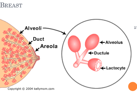
Mammary Glands
Modified sweat glands that produce milk (lactation).
Amount of adipose determines size of breast.
Milk-secreting glands open by lactiferous ducts at the nipple.
Areola is pigmented area around nipple.
Suspensory ligaments suspend breast from deep fascia of pectoral muscles (aging & Cooper’s droop).
Mammary line is a thickened ridge of embryonic tissue that extends from the axilla to the groin.
.jpg)
Prolactin from the pituitary gland stimulates the synthesis of milk.
Oxytocin from the posterior pituitary gland stimulates milk ejection.
Lymphatic drainage— lymph nodes draining the breast are located in the axilla.
.jpg)
Unlike males, who are able to produce sperm cells throughout their reproductive lives, females produce a finite number of egg cells.
During early fetal development germ cells migrate into the ovaries and differentiate into oogonia.
Oogenesis
Before birth
During fetal development, oogonia (stem cells) divide by mitosis to make primary oocytes.
By fifth month there are about 7 million primary oocytes, but most will degenerate during the next 2 months.
Primary oocytes begin meiosis and stop in prophase I until puberty.
Primordial follicles: Support cells that surround the oocyte in the ovary.
1—2 million present at birth.
400,000 remain at puberty.
After birth
Each month, hormones cause several follicles to develop, which triggers the primary oocyte to resume meiosis I.
Polar bodies: When the cell divides, all the cytoplasm and organelles stay with one of the new cells, the other cell is just DNA, and is called a polar body and is discarded.
Secondary oocyte: The stage at which ovulation occurs.
The secondary oocyte begins meiosis II, but stops in metaphase II.
The secondary oocyte is ovulated.
Meiosis II is completed only if it is fertilized.
.jpg)
Ovaries
Each follicle consists of an immature egg called an oocyte
Cells around the oocyte are called:
- Follicle cells (one cell layer thick). They are stimulated to mature by FSH from the pituitary gland.
- Granulosa cells (when more than one layer is present).
- Thecal cells: Cells in the ovarian stroma
Thecal & granulosa cells work together to produce estrogen.
A protective
Ovarian cycle
Monthly changes that occur in the ovary during a woman’s reproductive life.
Each month FSH stimulates primordial follicles to grow and mature (follicular phase).
Ovulation- release of the egg (LH).
In the luteal phase the corpus luteum produces progesterone that maintains uterine walls.
If fertilization does not occur, the corpus luteum degenerates, within 2 weeks into a mass of scar tissue called the corpus albicans.
.jpg)
Follicle Development
Primordial follicle: one layer of squamous-like follicle cells surrounds the oocyte.
Primary follicle: two or more layers of cuboidal granulosa cells enclose the oocyte.
Secondary follicle: has a fluid-filled space between granulosa cells that coalesces to form a central antrum.
Graafian follicle: secondary follicle at its most mature stage that bulges from the surface of the ovary.
Corpus luteum: ruptured follicle after ovulation.
.png)
.png)
.png)
.png)
.png)
Corpus luteum
- After ovulation, the remains of the follicle are transformed into a structure called the corpus luteum.
- If a pregnancy occurs, it produces progesterone to maintain the wall of the uterus during the early period of development.
Corpus albicans
- If fertilization does not occur, the corpus luteum will begin to break down about 2 weeks after ovulation.
- Degeneration occurs when fibroblasts enter the corpus luteum and a clump of scar tissue forms called the corpus albicans.
.jpg)
.jpg)
.jpg)
Practice Questions
Check how well you grasp the concepts by answering the following questions...
- This content is not available yet.
Send your comments, corrections, explanations/clarifications and requests/suggestions