What You Will Learn
After reading this note, you should be able to...
- This content is not available yet.
Read More 🍪
- It is an embryonal tumour.
- Most common primary ocular malignancy of childhood.
- It is a small round blue cell tumor arising from any of the layers of the retina.
- Usually unilateral (>75%).
- A relatively slow growing tumour.
- Late presentation is common, so blindness is a common presentation.
- Condition occurs globally.
- Incidence is about one per 16,000 – 18,000 live births globally.
- Worldwide, approximately 8,000 children develop retinoblastoma each year.
- Generally is the third or fourth commonest childhood malignancy in Nigeria after Burkitt's lymphoma and leukemia (nephroblastoma more common in some studies).
- May be heritable (about 40%) or non-heritable (about 60%).
- Equal sex predilection.
- Majority of cases are heritable.
- Mutation affecting the RB1 gene is implicated (germline or non-germline).
- Mutant allele is recessively inherited and requires a two-hit.
- Disease results from inactivation of the retinoblastoma gene (13q14).
- Encodes the retinoblastoma protein (pRb), a tumor suppressor protein that controls cell-cycle phase transition.
- Hereditary forms are usually bilateral and multifocal.
- Originates from the precursors of rods and cone cells of the retina arising from the posterior portions of the inner and outer nuclear layers.
- It forms nodules often with satellite seedlings.
- Tumour growth may be endophytic or exophytic (subretinal fluid and retinal detachment).
- 90% of cases occur before 5 years of age; most cases will occur before the 2nd year of life.
- Earlier presentation in the heritable form.
- May be detected incidentally.
- Leucokoria (white reflex or cat's eye reflex).
- Strabismus
- Pain
- Poor vision
- Orbital inflammation
- Pupillary irregularity
- Other features include nystagmus, failure to thrive, hyphema, proptosis, and fungating mass.
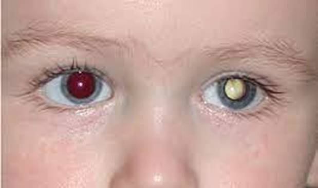
Leucokoria
×
![]()
Leucokoria
- Congenital cataract
- Retinopathy of prematurity
- Xerophthalmia
- Hyperplastic primary vitreous
- Coats disease
- Rhabdomyosarcoma
- Chloroma
- Neuroblastoma
- Retroorbital capillary hemangioma
| Stage | Characteristics |
|---|---|
| Stage I | Solitary or multiple tumours less than 4 disc diameter at or behind the equator of the eye |
| Stage II | Solitary or multiple tumours 4-10 disc diameter at or behind the equator of the eye |
| Stage III | Any lesion anterior to the equator, solitary tumours > 10 DD behind the equator |
| Stage IV | Multiple tumours greater than 10 DD, extending anterior to the ora serrata |
| Stage V | Massive tumours involving over half of the retina with vitreous seeding |
- Careful ophthalmologic examination
- May need to be done under general anaesthesia
- Orbital ultrasound scan
- Orbital radiographs: calcification in 75% of cases
- Cranial CT scan: To determine extent of tumour
- Cranial MRI: Define extent of tumour involvement including nerve involvement
- Treatment is determined by the size and location of the tumor.
- The primary goal is cure; the secondary goal is preserving vision.
- Unilateral: Unilateral enucleation of the eye
- Bilateral disease: Radiation to save less affected eye. Another option is chemoreduction in combination with focal therapy (laser photocoagulation or cryotherapy)
- Chemotherapy: Vincristine, etoposide, and carboplatin every 21-28 days for 6 months
- Enucleation: Removal of the entire globe and its intraocular contents, with preservation of all other periorbital and orbital structures
- Evisceration: Removal of the internal contents of the eye followed usually by placement of an orbital implant to replace the lost ocular volume
- Exenteration: Removal of the eye and its orbital contents including the extraocular muscles, orbital fat, nerves, lids, lashes, lachrymal gland, and occasionally brow
- Prognosis is good with early presentation.
- Most cases present late in this environment with poor prognosis for cure and preservation of vision.
Practice Questions
Check how well you grasp the concepts by answering the following questions...
- This content is not available yet.
Read More 🍪
Contributors
Jane Smith
She is not a real contributor.
John Doe
He is not a real contributor.
Send your comments, corrections, explanations/clarifications and requests/suggestions